|
Case Report
Mucinous adenocarcinoma in a young female: A case report of unusual-origin low-grade neoplasm presenting as retroperitoneal mass
1 Assistant Professor, Govt. College of Nursing, GSVM Medical College, Kanpur, Uttar Pradesh, India
2 Faculty of Nursing, Govt. College of Nursing, Sarojini Naidu Medical College, Agra, Uttar Pradesh, India
3 Dept. of Gastro Surgery, G.I. Oncology & HPB Surgery, CKS Hospital, Jaipur, Rajasthan, India
Address correspondence to:
Shashi Prakash
Assistant Professor, Govt. College of Nursing, GSVM Medical College, Kanpur, Uttar Pradesh 208002,
India
Message to Corresponding Author
Article ID: 100013C01SP2025
Access full text article on other devices

Access PDF of article on other devices

How to cite this article
Prakash S, Gundaiya S, Yadav L. Mucinous adenocarcinoma in a young female: A case report of unusual-origin low-grade neoplasm presenting as retroperitoneal mass. Edorium J Cancer 2025;9(2): 1–6.ABSTRACT
Introduction: Mucinous adenocarcinoma of the ovary is an uncommon type of epithelial ovarian cancer that predominantly occurs in women between their fourth and sixth decades of life. Presentation in younger women with atypical features, such as retroperitoneal cystic masses, is rare and will tend to delay diagnosis.
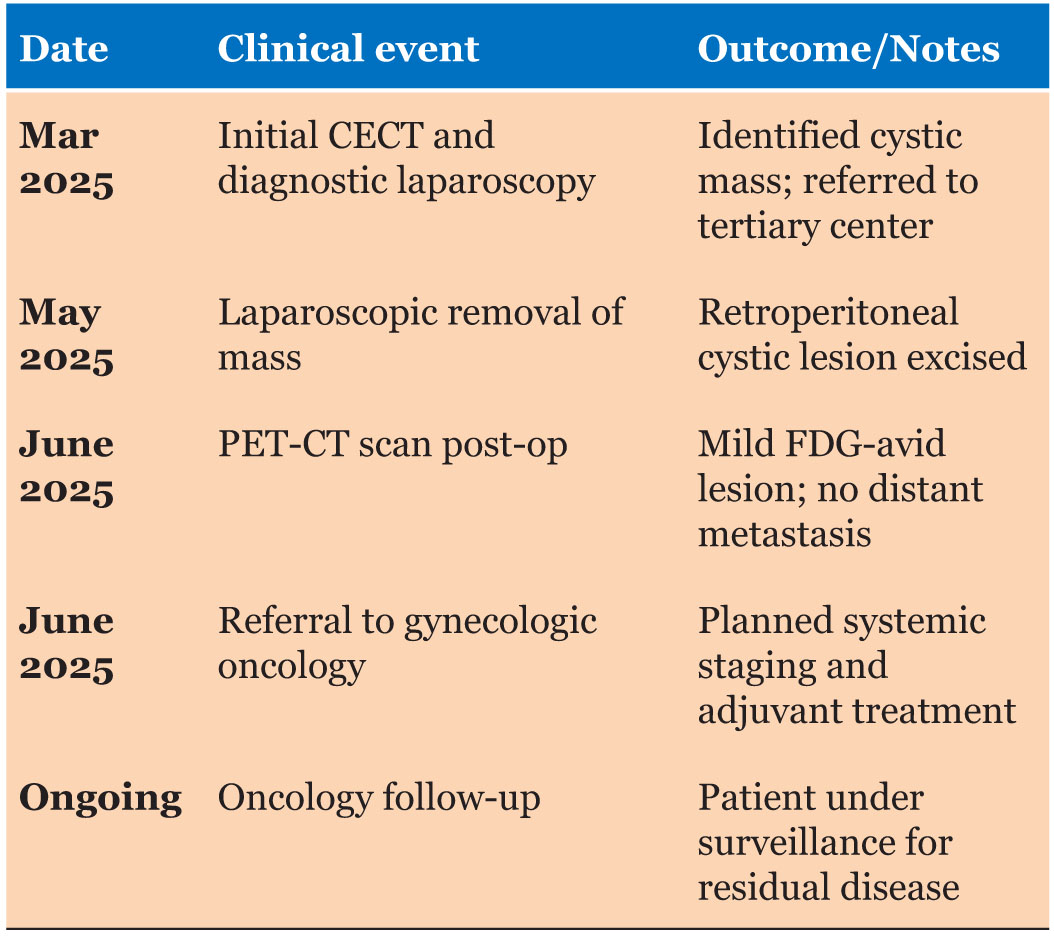
Case Report: After two caesarean sections, a 33-year-old multiparous woman (G2P2L2A0) experienced one to two months of right lower abdominal pain and swelling. A huge, multiloculated abdominopelvic cystic lesion on the right side was revealed by an imaging. The tumor was removed by laparoscopic surgery. Gross pathology showed an ovarian cyst with smooth walls measuring 22 × 14 × 10 cm. A mucinous cystadenoma was initially intimated by histopathological examination, but later examination and immunohistochemistry presented features consistent with a low-grade mucinous adenocarcinoma of probable ovarian origin. Immunostaining for PAX-8, WT-1, and CDX2 was negative, while that for CK7 and CA-125 was positive. In the absence of any suggestion of distant metastasis, postoperative positron emission tomography-computed tomography (PET-CT) demonstrated a moderately FDG-avid cystic mass in the right adnexa, suggesting persistent disease.
Conclusion: This case highlights the importance of an exhaustive immunohistochemical and histological evaluation in young women presenting with complex pelvic tumors. Mucinous adenocarcinoma should be identified at an early stage to guide treatment and improve prognosis. For optimal management, an imaging-based multidisciplinary approach with surgery and oncologic consultation is required.
Keywords: Computed tomography, Cystadenocarcinoma, Immunohistochemistry, Laparoscopy, Mucinous, Mucinous adenocarcinoma, Ovarian neoplasms, Tomography, Young adult
SUPPORTING INFORMATION
Author Contributions
Shashi Prakash - Substantial contributions to conception and design, Acquisition of data, Analysis of data, Interpretation of data, Drafting the article, Revising it critically for important intellectual content, Final approval of the version to be published
Shankar Gundaiya - Substantial contributions to conception and design, Acquisition of data, Drafting the article, Final approval of the version to be published
Lokesh Yadav - Substantial contributions to conception and design, Acquisition of data, Drafting the article, Final approval of the version to be published
Guaranter of SubmissionThe corresponding author is the guarantor of submission.
Source of SupportNone
Consent StatementWritten informed consent was obtained from the patient for publication of this article.
Data AvailabilityAll relevant data are within the paper and its Supporting Information files.
Conflict of InterestAuthors declare no conflict of interest.
Copyright© 2025 Shashi Prakash et al. This article is distributed under the terms of Creative Commons Attribution License which permits unrestricted use, distribution and reproduction in any medium provided the original author(s) and original publisher are properly credited. Please see the copyright policy on the journal website for more information.