Case Report
Mucinous adenocarcinoma in a young female: A case report of unusual-origin low-grade neoplasm presenting as retroperitoneal mass
1 Assistant Professor, Govt. College of Nursing, GSVM Medical College, Kanpur, Uttar Pradesh, India
2 Faculty of Nursing, Govt. College of Nursing, Sarojini Naidu Medical College, Agra, Uttar Pradesh, India
3 Dept. of Gastro Surgery, G.I. Oncology & HPB Surgery, CKS Hospital, Jaipur, Rajasthan, India
Address correspondence to:
Shashi Prakash
Assistant Professor, Govt. College of Nursing, GSVM Medical College, Kanpur, Uttar Pradesh 208002,
India
Access full text article on other devices

Access PDF of article on other devices
Article ID: 100013C01SP2025
doi: 10.5348/100013C01SP2025CR
How to cite this article
Prakash S, Gundaiya S, Yadav L. Mucinous adenocarcinoma in a young female: A case report of unusual-origin low-grade neoplasm presenting as retroperitoneal mass. Edorium J Cancer 2025;9(2): 1–6.ABSTRACT
Introduction: Mucinous adenocarcinoma of the ovary is an uncommon type of epithelial ovarian cancer that predominantly occurs in women between their fourth and sixth decades of life. Presentation in younger women with atypical features, such as retroperitoneal cystic masses, is rare and will tend to delay diagnosis.
Case Report: After two caesarean sections, a 33-year-old multiparous woman (G2P2L2A0) experienced one to two months of right lower abdominal pain and swelling. A huge, multiloculated abdominopelvic cystic lesion on the right side was revealed by an imaging. The tumor was removed by laparoscopic surgery. Gross pathology showed an ovarian cyst with smooth walls measuring 22 × 14 × 10 cm. A mucinous cystadenoma was initially intimated by histopathological examination, but later examination and immunohistochemistry presented features consistent with a low-grade mucinous adenocarcinoma of probable ovarian origin. Immunostaining for PAX-8, WT-1, and CDX2 was negative, while that for CK7 and CA-125 was positive. In the absence of any suggestion of distant metastasis, postoperative positron emission tomography-computed tomography (PET-CT) demonstrated a moderately FDG-avid cystic mass in the right adnexa, suggesting persistent disease.
Conclusion: This case highlights the importance of an exhaustive immunohistochemical and histological evaluation in young women presenting with complex pelvic tumors. Mucinous adenocarcinoma should be identified at an early stage to guide treatment and improve prognosis. For optimal management, an imaging-based multidisciplinary approach with surgery and oncologic consultation is required.
INTRODUCTION
Mucinous ovarian carcinoma (MOC) is an uncommon histologic type of epithelial ovarian cancer that accounts for approximately 3–5% of all primary ovarian cancers and about 10–15% of all mucinous ovarian tumors [1],[2]. Most mucinous tumors are either benign or borderline in type, with invasive carcinomas being significantly less frequent [3],[1]. The fifth to sixth decades of life are the peak ages of incidence, and less than 5% of MOC are diagnosed in women younger than 40 [4],[5]. This epidemiologic rarity is relevant, as it affects clinical suspicion and therapeutic decision in young patients [6],[7].
Large, unilateral, cystic, multilocular structures with smooth contours are characteristic on macroscopic appearance; expansile patterns of invasion, mucinous content, and calcification are distinguishing features [4]. Unlike these features, mucinous neoplasms of gastrointestinal (GI) origin are usually smaller in size, bilateral, exhibit nodularity or surface involvement, and often present as Krukenberg tumors with signet ring cells [8].
To separate primary MOC from metastatic mucinous carcinoma, immunohistochemistry (IHC) is necessary. With co-expression of CK7 being common and CK20 being focal to diffuse, MOC typically has a CK7+/CK20-variable profile. CDX2 tends to be low-to-moderately positive, whereas PAX8, WT1, and SATB2 are generally negative [9]. With great sensitivity, a panel of CK7, CK20, CDX2, PAX8, and SATB2 can differentiate primary ovarian from metastatic GI cancers [9].
Molecularly, MOCs frequently contain KRAS mutations (~46–64%), TP53 (~25–52%), and HER2 amplification (18–28%) and less frequently have changes in CDKN2A, ARID1A, and BRAF [1]. These genetic alterations provide potential treatment options in severe or recurrent disease as well as underpinning the distinctive pathophysiology of MOC.
Clinically, MOC typically manifests early; between 54% and 80% of patients are diagnosed as International Federation of Gynecology and Obstetrics (FIGO) stage I [4]. However, because of relative chemoresistance, advanced-stage illness has a poor prognosis; five-year survival drops from over 90% in early-stage patients to about 12% in advanced-stage disease. In young patients when fertility preservation is a clinical concern, this emphasizes the importance of early identification and the best possible surgical staging.
Here, we describe a 33-year-old woman who had low-grade mucinous adenocarcinoma, which began as an ovarian tumor that appeared as a cystic lesion in the right paracolic and retroperitoneal area. In addition to highlighting diagnostic difficulties, this case demonstrates how important multimodal evaluation—which includes imaging, histology, immunoprofiling, and molecular insights—is in directing the best course of action.
CASE REPORT
In May 2025, a 33-year-old woman (G2P2L2A0) arrived at the gastro-surgery department complaining of a palpable mass in the right iliac area, edema, and ongoing lower abdomen pain that had persisted for one to two months. She had never before experienced such a condition. Two full-term cesarean births in 2019 and 2023 were part of her obstetric history; both children were healthy and developed normally.
Clinical Examination
On examination, the patient was afebrile with stable vitals: Blood pressure (BP) 120/80 mmHg, pulse rate (PR) 76/min, Temp 98°F, SpO2 99%. Abdominal palpation revealed a firm, non-tender mass in the right lower quadrant.
Initial Imaging and erral
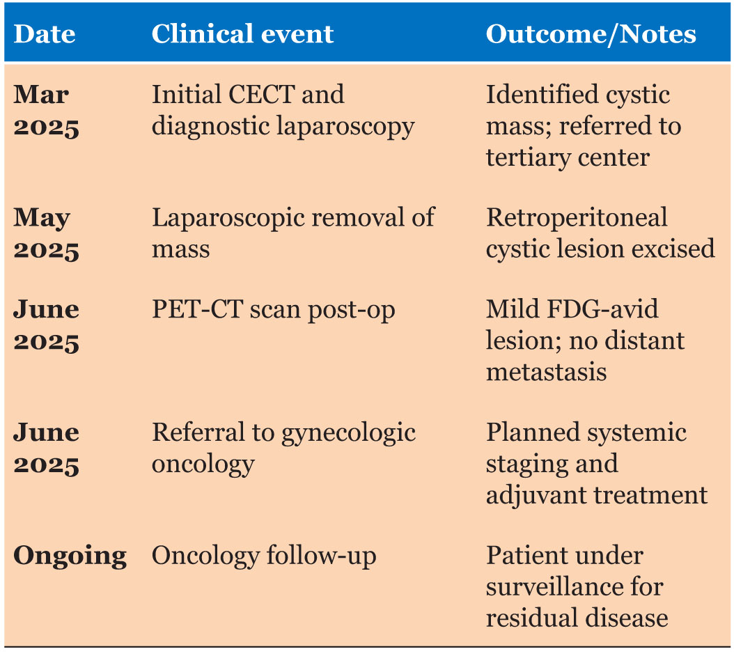
On March 2025, an external contrast-enhanced CT (CECT) scan showed a massive, distinct, multilocular cystic lesion in the right abdominopelvic area that measured 99 × 95 104 mm. The cyst had septations within it, nodular calcification in some locations, and had a thin wall. There were no mural nodules or post-contrast enhancement signs. The patient was erred to our hospital for final treatment following a diagnostic laparoscopy at a peripheral facility in view of these observations.
Surgical Procedure and Intraoperative Findings
On May 2025, the lump was removed laparoscopically while under general anesthesia. A sizable 15 × 15 cm cystic mass was discovered in the right paracolic gutter during surgery. It appeared to be retroperitoneal in origin and was not attached to the appendix or bowel.
Pathological Findings
Gross pathology
The removed specimen was a 22 × 14 × 10 cm ovarian cyst with a fallopian tube attached. The outside was smooth. Necrotic material was present in the multiloculated cyst on the cut segment. The fallopian tube looked remarkably normal.
Microscopic pathology
Histological analysis showed a multiloculated cyst with tiny to medium-sized nuclei and tall columnar epithelium lining it. Apical mucin was also prominent. There was no evidence of nuclear atypia or cellular stratification. Fibrocollagenous stroma was visible on the cyst wall. These findings are in keeping with a mucinous cystadenoma.
Immunohistochemistry (IHC)
On review and repeat staining, the tumor showed:
- CK7: Diffuse positive
- CK20: Focal dim positive
- PAX8, WT-1, ER, CDX2, SATB2: Negative
- CA-125: Patchy positive
- P53: Wild type pattern
The IHC profile was consistent with the diagnosis of mucinous cystadenocarcinoma, likely ovarian.
Multiple cysts and tubules lined by complicated columnar epithelium with nuclear pleomorphism, stratification, papillary structures, and intraluminal mucin pools with necrotic debris were observed upon a second study of paraffin-embedded sections (blocks C, D, and E). Low-grade mucinous adenocarcinoma was confirmed by these characteristics.
Biochemical and tumor marker profile
- CA-19.9: 41.4 U/mL (elevated)
- CA-125: Elevated
- CEA: 2.9 ng/mL (within normal range)
Positron Emission Tomography-Computed Tomography (PET-CT) Findings (June 2025)
There was no conclusive indication of distant metastases on postoperative PET-CT. In the right adnexal region, a weakly FDG-avid (SUVmax 1.5) ill-defined cystic lesion with internal septations was observed; this lesion is probably a surgical alteration or a lingering disease. The areas of the body that were inspected showed no additional FDG-avid lesions or lymphadenopathy. It was thought that breast FDG uptake might be lactational.
Whole-body coronal and sagittal PET-CT reconstruction showing a well-defined retroperitoneal mass arising near the left adnexal region, consistent with a mucinous adenocarcinoma of ovarian origin in a 33-year-old female patient. Metabolic activity is evident in the region of the lesion, suggestive of low-grade malignancy (Figure 1).
Coronal (left) and sagittal (right) images of this PET-CT scan show a retroperitoneal pelvic mass (white arrows) in a female patient, age 33, who has been diagnosed with mucinous adenocarcinoma, most likely of ovarian origin. With modest FDG avidity and a well-defined, hypodense appearance, the lesion suggests low-grade malignant activity. Its anatomical location and preserved fat planes confirm the diagnosis of a non-infiltrative neoplasm. Identification of the origin, planning of surgery, and confirmation of postoperative residual disease were all facilitated by the imaging.
Final Prognosis
Very likely to be ovarian in origin, mucinous adenocarcinoma presents as a retroperitoneal cystic tumor with low-grade malignancy histopathological features.
Management and Follow-up
On June 2025, the patient was erred to the gynecologic oncology service for adjuvant therapy and close follow-up after discharge in stable condition. Multidisciplinary tumor board discussions resulted in planning for further surgical staging and systemic treatment for the possibility of residual disease in the right adnexa.

DISCUSSION
Within the category of epithelial ovarian cancers, mucinous ovarian carcinoma (MOC) is an uncommon phenomenon that accounts for roughly 3–5% of ovarian malignancies and 10–15% of mucinous ovarian tumors, the majority of which are benign or borderline [6]. MOC in females under 40, especially those in their 20s or 30s, is uncommon, in contrast to the usual appearance in older women. It is uncommon, but because of its unique behavior and therapeutic implications, it deserves managerial attention.
Mucinous ovarian carcinoma is considered to take a stepwise pathway model, developing from benign mucinous cystadenomas to borderline tumors and finally to invasive carcinoma. This development is initiated by initial KRAS mutations early on, followed by other alterations like TP53 mutations or HER2 amplification in high-grade lesions. This expansile invasion pattern and mucinous differentiation are typical of this pathway.
Histopathology and Tumor Progression
With increasing cytologic atypia and invasiveness, MOC often arises as a gradual evolution of benign or borderline cystadenomas to carcinoma [6]. Mucinous tumors are also unilocular or multilocular, filled with mucinous fluid, and increase to large dimensions—some of them over 20 cm [6]. Epithelial stratification, papillary patterns, expansile or infiltrating invasion, and intraluminal mucin pools are histological characteristics of low-grade cancer. In this case, the diagnosis of mucinous adenocarcinoma was confirmed by the demonstration of mucin pools, papillary appearance, and nuclear pleomorphism [10].
Differential Diagnosis: Distinguishing Primary from Metastatic
A mixture of clinical, histologic, and immunohistochemical features is required to distinguish primary MOC from metastatic mucinous carcinomas to the ovary, i.e., from the gastrointestinal or pancreatobiliary tracts, as they may mimic one another [8]. Whereas metastatic tumors tend to be bilateral, small, and composed of much external mucin, primary MOC tends to remain unilateral with much intracellular mucin [6].
Immunohistochemical profiling is a critical discriminator:
- CK7: Diffuse staining in ~90% of primary MOC [10].
- CK20: Focal or variable in MOC; diffuse positivity indicates colorectal or appendiceal metastasis [8].
- CDX2: Normally negative or focal in MOC, but strongly positive in lower GI neoplasms [8].
- PAX8 and WT-1: Frequently negative in MOC; positive in Müllerian-origin neoplasms [8].
- SATB2: Infrequently expressed in MOC but strongly positive in lower GI metastases [8].
This case’s IHC pattern—CK7 diffuse+, CK20 focal+, CDX2 negative, PAX8/WT 1/SATB2 negative—is highly suggestive of a primary ovarian origin. This type of pattern is consistent with reports of successful differentiation via a CK7/CK20/CDX2 panel [8].
Molecular Characteristics
Genetically, MOC often has KRAS mutations (30–60%), and may also have TP53 mutations, HER2 amplification, and rare changes in CDKN2A, ARID1A, and BRAF [2],[11],[12]. Approximately 20–30% of patients exhibit HER2 amplification, which has therapeutic significance; in certain cases, targeted treatments, such as trastuzumab, have proven beneficial [2].
In order to guide therapy, KRAS and HER2 status should be evaluated during adjuvant management, even though molecular data were unavailable for this patient.
Clinical behavior and prognosis
MOC stands out due to its early detection and poor response to conventional platinum-based treatment [6]. The majority of patients arrive at FIGO stage I, and over 90% of them survive for five years. However, chemo-resistance contributes to the poor results (~12% survival) of advanced-stage illness (III/IV). Theore, a timely and precise diagnosis is essential. In young adults and adolescents, fertility-sparing surgery is frequently contemplated. According to reports, unilateral oophorectomy and surgical staging for early-stage illness can result in acceptable oncologic results [6]. Conservative surgical methods when appropriate were supported by a multicenter U.S. trial that compared fertility-sparing and radical surgery for stage I epithelial ovarian tumors, including mucinous subtype, and found no difference in 10-year survival (˜88%) [6].
Role of imaging and tumor markers
CA 19-9, and carcinoembryonic antigen (CEA) are examples of tumor markers that can help in diagnosis and follow-up. While CA 19-9 and CEA may indicate GI-origin tumors, CA 125 is more frequently increased in ovarian-origin mucinous tumors [6]. Along with CEA within the normal range, our high CA 125 and CA 19-9 additionally supported ovarian origin.
Computed tomography and PET CT are imaging modalities that assist in residual disease detection and staging. In this case, localized disease was suggested by PET CT, which detected a small FDG-avid adnexal lesion but no distant metastases. PET CT makes postoperative evaluation easier, but MRI may be needed to assess brain metastases.
Management implications
The primary course of treatment continues to be surgical excision combined with thorough staging. It is recommended to do lymphadenectomy and omentectomy because of the low proliferative nature of mucinous adenocarcinoma. When pregnancy is desired and the disease is in stage I low grade, fertility-sparing methods are appropriate.
For stage IC or above, adjuvant chemotherapy is usually advised. But because MOC is less responsive to chemotherapy, there is interest in other approaches. Women with HER2-amplified malignancies may benefit from targeted therapy, especially anti-HER2. Immunotherapies, PARP inhibitors, and MEK inhibitors are being investigated in clinical studies for mucinous ovarian cancer.
Review of Literature
Earlier series and case reports have established the rarity of mucinous ovarian carcinoma in females below the age of 35. One review reported that less than 5% of all mucinous ovarian carcinomas are detected below the age of 40. Seidman et al. have also highlighted in a study the difficulty in differentiating primary ovarian mucinous carcinoma from metastatic lesions with IHC, with the accuracy increasing when a panel for diagnosis is employed. Fertility-sparing operations have been reported to result in favorable results in stage I low-grade cases [2],[7],[11],[12].
Follow-up and Surveillance
It is crucial to conduct thorough follow-up with clinical examinations, imaging, and tumor marker monitoring due to the danger of late recurrence, even after fertility preservation. If it is practical, salvage surgery can be used to treat recurrence. It is frequently advised to conduct surveillance for up to ten years [7].
CONCLUSION
This case demonstrates a rare presentation of low-grade mucinous adenocarcinoma in a young woman, mimicking a retroperitoneal cystic lesion. It underscores the importance of maintaining a broad differential diagnosis when evaluating adnexal masses in reproductive-age patients. Definitive diagnosis required integrated imaging, surgical excision, and immunohistochemical profiling, which supported a primary ovarian origin. PET-CT was valuable in postoperative surveillance. Timely intervention and multidisciplinary coordination were essential in guiding management. Given the tumor’s limited response to conventional chemotherapy, further research into molecular profiling and targeted therapies is warranted.
REFERENCES
1.
Babaier A, Ghatage P. Mucinous cancer of the ovary: Overview and current status. Diagnostics (Basel) 2020;10(1):52. [CrossRef]
[Pubmed]

2.
Köbel M, Kalloger SE, Boyd N, McKinney S, Mehl E, Palmer C, et al. Ovarian carcinoma subtypes are different diseases: Implications for biomarker studies. PLoS Med 2008;5(12):e232. [CrossRef]
[Pubmed]
3.
Khanna SR, Nguyen B, Charif M. Early-stage mucinous ovarian adenocarcinoma with extensive clotting in a previously healthy young female patient: An uncommon presentation of a relatively uncommon disease. Am J Case Rep 2017;18:669–73. [CrossRef]
[Pubmed]
4.
Zhu S, Yao M, Xiong L, Tan B. Giant mucinous cystadenocarcinoma of ovary in a young woman: A case report and review of literature. Front Oncol 2024;14:1377042. [CrossRef]
[Pubmed]
5.
Feinstein L. Letter to the editor regarding “Giant mucinous cystadenocarcinoma of ovary in a young woman: A case report and review of literature”. Front Oncol 2024;14:1443334. [CrossRef]
[Pubmed]
6.
Agarwal N, Garg R, Gandhi G, Goodman A. Mucinous ovarian malignancy in a young woman: A case report. Clin Case Rep J 2021;2(5):1–5.
7.
Heller D, Krestel R, Ohler U, Vingron M, Marsico A. ssHMM: Extracting intuitive sequence-structure motifs from high-throughput RNA-binding protein data. Nucleic Acids Res 2017;45(19):11004–18. [CrossRef]
[Pubmed]
8.
Dundr P, Singh N, Nožičková B, Němejcová K, Bártů M, Stružinská I. Primary mucinous ovarian tumors vs. ovarian metastases from gastrointestinal tract, pancreas and biliary tree: A review of current problematics. Diagn Pathol 2021;16(1):20. [CrossRef]
[Pubmed]
9.
Strickland S, Wasserman JK, Giassi A, Djordjevic B, Parra-Herran C. Immunohistochemistry in the diagnosis of mucinous neoplasms involving the ovary: The added value of SATB2 and biomarker discovery through protein expression database mining. Int J Gynecol Pathol 2016;35(3):191–208. [CrossRef]
[Pubmed]
10.
Hamza HK, Yaqoob N, Galaal KA, Zribi A, Al-Haddabi I. Mucinous carcinoma of ovary in a 15-year-old girl, a rare case report and literature review. Turk Patoloji Derg 2024. [CrossRef]
[Pubmed]
11.
Anglesio MS, Kommoss S, Tolcher MC, Clarke B, Galletta L, Porter H, et al. Molecular characterization of mucinous ovarian tumours supports a stratified treatment approach with HER2 targeting in 19% of carcinomas. J Pathol 2013;229(1):111–20. [CrossRef]
[Pubmed]
12.
Ryland GL, Hunter SM, Doyle MA, Caramia F, Li J, Rowley SM, et al. Mutational landscape of mucinous ovarian carcinoma and its neoplastic precursors. Genome Med 2015;7(1):87. [CrossRef]
[Pubmed]
SUPPORTING INFORMATION
Author Contributions
Shashi Prakash - Substantial contributions to conception and design, Acquisition of data, Analysis of data, Interpretation of data, Drafting the article, Revising it critically for important intellectual content, Final approval of the version to be published
Shankar Gundaiya - Substantial contributions to conception and design, Acquisition of data, Drafting the article, Final approval of the version to be published
Lokesh Yadav - Substantial contributions to conception and design, Acquisition of data, Drafting the article, Final approval of the version to be published
Data Availability StatementThe corresponding author is the guarantor of submission.
Consent For PublicationWritten informed consent was obtained from the patient for publication of this article.
Data AvailabilityAll relevant data are within the paper and its Supporting Information files.
Competing InterestsAuthors declare no conflict of interest.
Copyright© 2025 Shashi Prakash et al. This article is distributed under the terms of Creative Commons Attribution License which permits unrestricted use, distribution and reproduction in any medium provided the original author(s) and original publisher are properly credited. Please see the copyright policy on the journal website for more information.